This is an accordion element with a series of buttons that open and close related content panels.
MASSIVE RETINAL GLIOSIS. A REACTIVE PROLIFERATION OF MÜLLER CELLS
Michael Nork // Publications // Sep 01 1986
PubMed ID: 3092790
Author(s): Nork TM, Ghobrial MW, Peyman GA, Tso MO. Massive retinal gliosis. A reactive proliferation of Müller cells. Arch Ophthalmol. 1986 Sep;104(9):1383-9. PMID 3092790
Journal: Archives Of Ophthalmology (Chicago, Ill. : 1960), Volume 104, Issue 9, Sep 1986
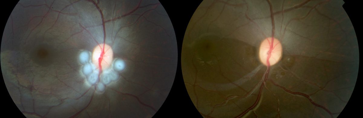
Both Müller cells and astrocytes have been implicated in the dispute over the histogenesis of massive retinal gliosis. We studied three cases of massive retinal gliosis by light and electron microscopy and immunocytochemistry. Spindle fibrillary glial cells were joined by zonulae adherentes resembling those of the external limiting membrane of the retina. Furthermore, these cells produced a continuous basement membrane around an extracellular space filled with fine filaments, which was highly suggestive of vitreous cavity. In the proliferating cells, immunoperoxidase technique disclosed the presence of carbonic anhydrase isoenzyme C, characteristically found only in Müller cells. The glial cells in the preretinal membrane away from the gliotic nodule showed similar characteristics. We concluded that both the nodule of massive retinal gliosis and the associated preretinal glial membrane resulted from the proliferation and migration of Müller cells.
SWELLING AND LOSS OF PHOTORECEPTORS IN CHRONIC HUMAN AND EXPERIMENTAL GLAUCOMAS
James Verhoeve // Matthew Davis // Michael Nork // Nickells Lab // Publications // Feb 01 2000
PubMed ID: 10676789
Author(s): Nork TM, Ver Hoeve JN, Poulsen GL, Nickells RW, Davis MD, Weber AJ, Vaegan, Sarks SH, Lemley HL, Millecchia LL. Swelling and loss of photoreceptors in chronic human and experimental glaucomas. Arch Ophthalmol. 2000 Feb;118(2):235-45. PMID 10676789
Journal: Archives Of Ophthalmology (Chicago, Ill. : 1960), Volume 118, Issue 2, Feb 2000
OBJECTIVE To determine whether outer retinal changes occur in chronic, presumed primary open-angle glaucoma (POAG).
METHODS The outer retinas from 128 human eyes with a diagnosis of chronic glaucoma (presumably POAG in most cases) and 90 control eyes were examined histologically by 3 masked observers for photoreceptor swelling and loss. Retinas from 9 rhesus monkeys with glaucoma induced experimentally by laser trabecular destruction were compared with 7 fellow (control) eyes. The mean pressure elevations in the eyes with laser trabecular destruction ranged from 26.6 to 53.6 mm Hg with durations varying from 7 to 33 weeks.
RESULTS Swelling of the red- and green-sensitive cones was observed in a statistically significantly greater proportion of human eyes with presumed POAG compared with the control eyes. Patchy loss of red/green cones and rods was also found in some of the glaucomatous retinas. In a subset of the human eyes with end-stage disease, cone swelling was a variable finding. Although no photoreceptor loss was found in the 9 monkey eyes with experimental glaucoma, 8 had swelling of their red/green cones that was remarkably similar to that seen in the human eyes. Swelling was not present in any of the control monkey eyes.
CONCLUSIONS The photoreceptors are affected by chronically elevated intraocular pressure.
CLINICAL RELEVANCE These findings may explain some of the abnormalities of color vision and the electrophysiological effects that have been observed in patients with POAG.
REGIONAL CHOROIDAL BLOOD FLOW AND MULTIFOCAL ELECTRORETINOGRAPHY IN EXPERIMENTAL GLAUCOMA IN RHESUS MACAQUES
Michael Nork // Publications // Nov 04 2014
PubMed ID: 25370515
Author(s): Nork TM, Kim CB, Munsey KM, Dashek RJ, Hoeve JN. Regional choroidal blood flow and multifocal electroretinography in experimental glaucoma in rhesus macaques. Invest Ophthalmol Vis Sci. 2014 Nov 4;55(12):7786-98. doi: 10.1167/iovs.14-14527. PMID 25370515
Journal: Investigative Ophthalmology & Visual Science, Volume 55, Issue 12, Nov 2014
PURPOSE To test a hypothesis of regional variation in the effect of experimental glaucoma on choroidal blood flow (ChBF) and retinal function.
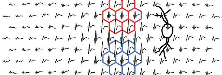
METHODS Five rhesus macaques underwent laser trabecular destruction (LTD) to induce elevated intraocular pressure (IOP). Intraocular pressures were elevated for 56 to 57 weeks. Multifocal electroretinographic (mfERG) and multifocal visual evoked cortical potential (mfVEP) testing were performed at regular intervals before and during the period of IOP elevation. At euthanasia, the IOP was manometrically controlled at 35 (experimentally glaucomatous eye) and 15 (fellow control eye) mm Hg. Fluorescent microspheres were injected into the left ventricle. Regional ChBF was determined.
RESULTS All of the experimentally glaucomatous eyes exhibited supranormal first-order kernel (K1) root mean square (RMS) early portions of the mfERG waveforms and decreased amplitudes of the late waveforms. The supranormality was somewhat greater in the central macula. Second-order kernel, first slice (K2.1) RMS mfVEP response was inversely correlated (R(2) = 0.97) with axonal loss. Total ChBF was reduced in the experimentally glaucomatous eyes. The mean blood flow was 893 ± 123 and 481 ± 37 μL/min in the control and glaucomatous eyes, respectively. The ChBF showed regional variability with the greatest proportional decrement most often found in the central macula.
CONCLUSIONS This is the first demonstration of globally reduced ChBF in chronic experimental glaucoma in the nonhuman primate. Both the alteration of mfERG waveform components associated with outer retinal function and the reduction in ChBF were greatest in the macula, suggesting that there may be a spatial colocalization between ChBF and some outer retinal effects in glaucoma.
Copyright 2014 The Association for Research in Vision and Ophthalmology, Inc.
FUNCTIONAL AND ANATOMIC CONSEQUENCES OF SUBRETINAL DOSING IN THE CYNOMOLGUS MACAQUE
James Verhoeve // Michael Nork // Publications // Richard Dubielzig // Jan 01 2012
PubMed ID: 21911651
Author(s): Nork TM, Murphy CJ, Kim CB, Ver Hoeve JN, Rasmussen CA, Miller PE, Wabers HD, Neider MW, Dubielzig RR, McCulloh RJ, Christian BJ. Functional and anatomic consequences of subretinal dosing in the cynomolgus macaque. Arch Ophthalmol. 2012 Jan;130(1):65-75. doi: 10.1001/archophthalmol.2011.295. Epub 2011 Sep 12. PMID 21911651
Journal: Archives Of Ophthalmology (Chicago, Ill. : 1960), Volume 130, Issue 1, Jan 2012
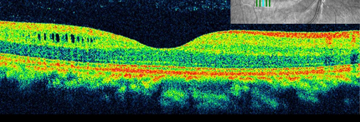
OBJECTIVE To characterize functional and anatomic sequelae of a bleb induced by subretinal injection.
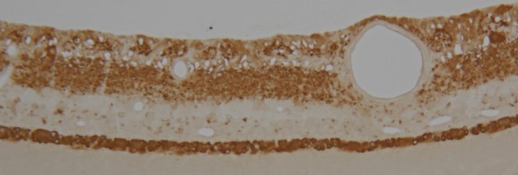
METHODS Subretinal injections (100 μL) of balanced salt solution were placed in the superotemporal macula of 1 eye in 3 cynomolgus macaques. Fellow eyes received intravitreal injections (100 μL) of balanced salt solution. Fundus photography, ocular coherence tomography, and multifocal electroretinography were performed before and immediately after injection and again at intervals up to 3 months postinjection. Histopathologic analyses included transmission electron microscopy and immunohistochemistry for glial fibrillary acidic protein, rhodopsin, M/L-cone opsin, and S-cone opsin.
RESULTS Retinas were reattached by 2 days postinjection (seen by ocular coherence tomography). Multifocal electroretinography waveforms were suppressed post-subretinal injection within the subretinal injection bleb and, surprisingly, also in regions far peripheral to this area. Multifocal electroretinography amplitudes were nearly completely recovered by 90 days. The spectral-domain ocular coherence tomography inner segment-outer segment line had decreased reflectivity at 92 days. Glial fibrillary acidic protein and S-cone opsin staining were unaffected. Rhodopsin and M/L-cone opsins were partially displaced into the inner segments. Transmission electron microscopy revealed disorganization of the outer segment rod (but not cone) discs. At all postinjection intervals, eyes with intravitreal injection were similar to baseline.
CONCLUSIONS Subretinal injection is a promising route for drug delivery to the eye. Three months post-subretinal injection, retinal function was nearly recovered, although reorganization of the outer segment rod disc remained disrupted. Understanding the functional and anatomic effects of subretinal injection is important for interpretation of the effects of compounds delivered to the subretinal space.
CLINICAL RELEVANCE Subretinal injection is a new potential route for drug delivery to the eye. Separating drug effects from the procedural effects is critical.